Do you want to know what a vertebral column is? Where is it located in the body and what is the importance of vertebrae? What are the shape and articulating surfaces and prominent features of the vertebrae? What is the anatomy of the vertebral column? Don’t worry, we will provide you with complete guidance. Please read this article carefully which will help you to understand the vertebral column and its unique features.
Consider the vertebral column, the body’s longitudinally oriented core skeletal column. In addition to supporting the skull, pectoral girdle, upper limbs, and thoracic cage, it conveys body weight to the lower extremities via the pelvic girdle. The spinal cord, the roots of the spinal nerves, and the meninges surrounding the spinal column are all protected by a hollow within the vertebral column. This depression is home to all of these structures.
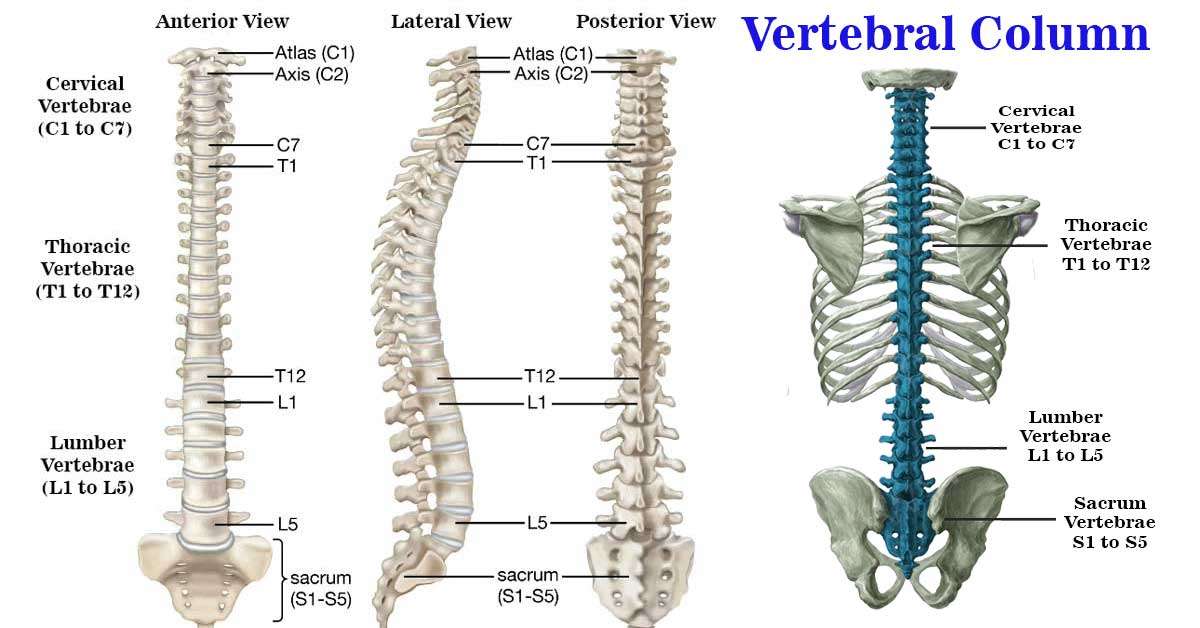
Seven cervical, twelve thoracic, five lumbar, five sacral (which, when joined, form the sacrum), and four coccygeal vertebrae comprise the vertebral column (the lower three are commonly fused). The vertebral column has great motion due to the numerous joints connecting the unique vertebral segments.
The Vertebrae
Each region’s vertebrae have unique characteristics that set them apart from vertebrae from other regions. Conversely, the essential structural arrangement of each vertebra is identical. The front of a typical vertebra consists of a round or spherical body, whereas the rear consists of an arch. They encompass a passageway known as the vertebral foramen.
Structure of Vertebral Column
The vertebral foramina of an articulated skeleton are positioned to form the vertebral canal, a continuous passageway. Along this canal, the spinal cord and its coverings are conveyed. A pair of cylindrical pedicles form the sides of the vertebral arch, while a pair of flattened laminae complete the posterior half of the arch. A pair of laminae connect the pedicles. The vertebral arch develops seven processes, including one spinous, two transverse, and four articular processes.
The spinous process, often known as the spine, covers posteriorly from the junction of the two laminae. From the intersection of the laminae and pedicles, the transverse processes extend in a lateral direction. Levers are the spinous and transverse processes and the attachment points of the muscles and ligaments. The total number of articular processes is four, with two superior and two inferior processes placed vertically. Their articular surfaces, often facets, are coated in hyaline cartilage and originate at the intersection of the laminae and pedicles.
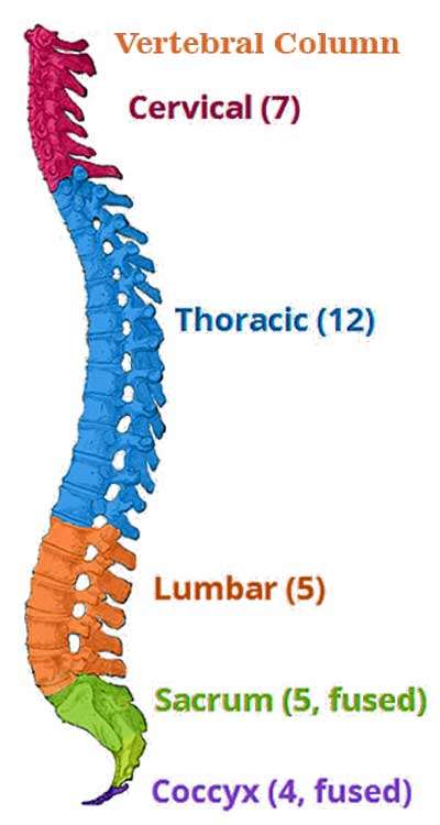
Two synovial joints are formed when the two superior articular processes of one vertebral arch come into contact with the two inferior articular processes of the arch above it. Two synovial joints are generated when the two inferior articular processes of the vertebral arch connect with the two superior articular processes of the arch below. Thus, each vertebra has a total of four synovial articular joints.
The superior and inferior vertebral notches are the notches on the pedicles located on the superior and inferior pedicle borders. The intervertebral foramen is formed on either side of an articulated skeleton when one vertebra’s prime notch aligns with the vertebra’s inferior notch above it. These foramina allow the transmission of blood vessels and spinal neurons. Before the anterior and posterior nerve roots of a spinal nerve may shed their meningeal coverings and form segmental spinal nerves, they must first rejoin within these foramina.
The Vertebral arch
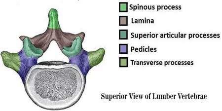
The vertebral arch comprises the lateral and posterior surfaces of each vertebra. The vertebral arch creates the vertebral foramen, a bone-enclosed opening resulting from the vertebral body and arch union. When the foramina of each vertebra align, they create the vertebral canal, which protects and shields the spinal cord.
Each vertebral arch features several bony prominences that serve as attachment points for several muscles and ligaments, including the following:
- Spinous processes: Each vertebra contains one spinous process centered posteriorly near the arch.
- Transverse processes — two transverse processes extend laterally and posteriorly, one on each side of the vertebral body. Both sides of the vertebral bodies include these features. Transverse processes of the thoracic vertebrae generate the joints that connect to the ribs.
- Pedicles connect the transverse processes to the vertebral body.
- The lamina, which joins the transverse and spinous processes, is a structural component.
- Articular processes form the joints between a vertebra and its anterior and posterior counterparts. The articular processes are located at the junction of the laminae and pedicles.
Atypical Vertebra
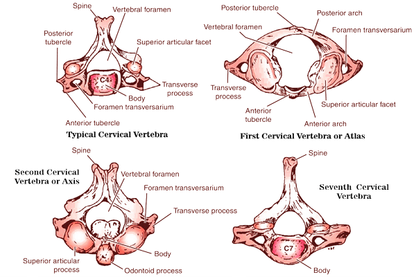
The first, second, and seventh cervical vertebrae are atypical, as are the first, tenth, eleventh, and twelfth thoracic vertebrae. The atlas, commonly known as the first cervical vertebra, is devoid of a body and spinous process. Instead, it is flanked on both sides by arches. It features articular surfaces on both its inferior and superior surfaces for articulation with the C2 vertebra and the occipital condyles of the skull, respectively (atlanto-occipital joints).
Each of its sides also possesses a lateral mass (atlantoaxial joints). The axis, also known as the second cervical vertebra, has an odontoid process that extends upward from the body’s superior surface. This operation is referred to as dens. The dens show the area of the atlas body that initially merged with the axis body in the early stages of development.
The name “vertebra prominens,” which refers to the seventh cervical vertebra, comes from the fact that it has the longest spinous process of any cervical vertebra, and that process is not bifid. Unlike the massive transverse process, the foramen transversarium, which transmits the vertebral vein or veins, is rather small. Variations in the rib attachments to the bodies and transverse processes of the first, tenth, eleventh, and twelfth thoracic vertebrae account for their atypical appearance.
Sacrum
The sacrum comprises five primordial vertebrae fusing to form a wedge-shaped bone. The anterior surface of the sacrum is concave. The fifth lumbar vertebra can articulate with the bone’s upper border or base. The coccyx and the narrow inferior border, often known as the apex, articulate. The sacroiliac joints are formed when the sacrum and the two iliac bones articulate laterally to form the sacrum.
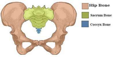
The anterior and upper edges of the first sacral vertebra project forward at the posterior limit of the pelvic inlet. The technical terminology for this sacral protuberance is employed. Obstetricians pay special attention to the sacral promontory when determining the size of a woman’s pelvis due to its vital role in reproduction.
After passing through the sacrum, the vertebral canal subsequently converts into the sacral canal. The sacral canal contains the cauda equina, composed primarily of the anterior and posterior roots of the sacral and coccygeal spinal nerves and fibro-fatty tissue. Moreover, it includes the entire subarachnoid space between the second sacral vertebrae and its lowest point.
The sacral gap develops when the laminae of the fifth sacral vertebra and occasionally those of the fourth fail to align to form a complete arch in the midline. Sacral hiatus is distinguished by the absence of a bony cap at the inferior posterior end of the sacral canal, which permits unrestricted access to the canal.
Each of the anterior and posterior surfaces of the sacrum has four foramina through which the anterior and posterior rami of the upper four sacral nerves exit (anterior and posterior sacral foramina). These foramina are positioned in the front and back of the sacrum. Sacralization of the L5 vertebra refers to the process through which the fifth lumbar vertebra may fuse with the sacrum. Typically, this is partial and may cover a single side.
In contrast, the first sacral vertebra may remain entirely or partially distinct from the sacrum and resemble the sixth lumbar vertebra (lumbarization of the S1 vertebra). It is conceivable for the laminae and spines of the sacral canal to fail to develop, resulting in a significant portion of the sacral canal’s posterior wall being absent.
Coccyx
Usually, four vertebrae fuse to form the coccyx, a tiny triangular bone that articulates with the lower end of the sacrum at its base. Typically, the first coccygeal vertebra does not merge with the second, or the fusion is just partial. The coccyx can have three or five vertebrae, depending on the individual. The first coccygeal vertebra may be a distinct entity.
Typically, the free vertebra expands anteriorly and downward from the apex of the sacrum when this illness is present. An alternative term for this condition is an anterior protrusion. For correctly reading radiographs and recognizing the locations of bony pathologic characteristics concerning soft tissue injuries, prior knowledge of the basic anatomy of the vertebral column is essential.
Functions of Vertebral Column
The vertebral column serves four primary purposes:
Protection: Within the spinal canal, it encloses and protects the spinal cord.
Support: Bears the body’s weight above the pelvis.
Axis: An axis forms the central axis of the body.
Movement: Participates in both posture and movement.
Curves in Vertebral Columns
In the standing position of an adult, the spinal column forms four unique regional curvatures in the sagittal plane. The cervical and lumbar curves feature posterior concavities, whereas the thoracic and sacrococcygeal curves have anterior concavities. The curves align the body’s center of gravity with the pelvis when regarded as a whole. The body can then maintain an upright posture because the head is properly balanced atop the spinal column, offsetting the weight of the thoraco-abdominal viscera and allowing for an upright stance.
During the early stages of development, the fetal spinal column contains only one continuous anterior concavity. As growth continues, the lumbosacral angle will eventually form at the junction of the L5 and S1 vertebrae. Hence, two anteriorly concave curves will be produced. At birth, the infant holds their head above the spinal column, lifting it above the level of the birth canal, resulting in the formation of the cervical curve.
The lumbar curve develops as a baby nears the end of their first year and begins to sit up and stand straight for the first time. As the thoracic and sacrococcygeal curves retain their original prenatal anterior concavities, they are primary curvatures. Secondary curvatures refer to the cervical and lumbar curves. It results from posterior concavities and the postnatal development of the cervical and lumbar curves. As a result of the development of secondary curves, the geometry of the vertebral bodies and intervertebral discs is altered.
Adult females have a more significant natural curve in their lower back than males. To maintain their center of gravity, as the size and weight of the fetus continue to increase, women frequently develop a greater lumbar concavity later in pregnancy. It is due to the continuous development of the fetus. As people age, the intervertebral discs begin to degrade, resulting in the vertebral column reverting to having a continuous anterior concavity. It will result in a diminution of height.
Late adolescent children with mild lateral spinal bends in the thoracic area are uncommon. It is extremely typical and frequently occurs when one of the upper limbs is favored over the other. For example, right-handed individuals typically have a slight convexity on the right side of their thoracic region. On either side of this curve are minor compensatory curves that go in the opposite direction.
Anomalous Vertebral Column Curves
Kyphosis is caused by a physical structural change in the vertebral bodies or intervertebral discs, which causes a considerable exaggeration of the thoracic curve of the vertebral column. In situations of disease-based kyphosis, the vertebral column becomes severely angular due to illnesses such as crush fractures or tuberculous-caused vertebral body loss. Due to osteoporosis, intervertebral disc degeneration, significant weakening of the intrinsic back muscles, and all three, age-related senile kyphosis can affect the cervical, thoracic, and lumbar sections of the spinal column. Long study hours or work at a low desk can result in a less severe, softly curving upper thoracic area in young persons with poor muscular tone. A “round-shouldered” condition may or may not be classified as kyphosis.
Lordosis is an exaggerated version of either of the secondary spinal curvatures. Although there may be considerable cervical curvature, the lumbar curve is usually emphasised. Lumbar lordosis can be induced by a vertebral column problem like spondylolisthesis or by an increase in the weight of the abdominal contents, such as with a gravid uterus or a big ovarian tumour. Furthermore, lumbar lordosis is likely a thoracic kyphosis or a hip joint issue that needs postural support (e.g., congenital dislocation).
Scoliosis is a lateral displacement of the spinal column that usually involves malrotation. There are different possible explanations for this availabe, which is most commonly found in the thoracic area. Scoliosis can be caused by poliomyelitis-related muscle paralysis or a congenital hemivertebra. Scoliosis is often compensatory and can be caused by a short leg or a hip issue. Scoliosis is more common in women, and the condition’s onset is directly tied to the adolescent growth spurt. Yet, most scoliosis cases are idiopathic (i.e., of unknown cause).

